Webbing of the lateral canthus (where outer upper and lower lids meet) is called a canthal web. Normally the lateral canthus consists of an almond shaped angle. When webbing occurs, skin obscures the angle and the canthal angle resembles a duck’s foot which is normally webbed. This causes both aesthetic disfigurement (it is not attractive), and a functional deficit (the web blocks vision on side gaze).
The best way to treat a canthal web is to avoid allowing it to develop. These webs occur in a variety of situations. I have had them sent to me form correction when:
1. Upper and lower lid surgery has been performed together and the outer portions of the upper and lower lid incision are too close to each other.
2. When a canthoplasty (lower lid tightening) has been added to surgery
3. When an infection has occurred after surgery
4. When a hematoma (bleed) has occurred after surgery
Canthal webs occur because there is a relative deficiency of skin in the vertical as compared to the horizontal plane. They are surgically corrected by a variety of micro-skin flaps. Most plastic surgeons avoid addressing canthal webs as they are difficult to correct and can lead to significant scaring. I have been referred numerous webs over the years and have developed a reliable means of correcting them. I recently published a peer reviewed manuscript titled “Cicatricial Canthal Webs.” It will appear in our society journal “Ophthalmic Plastic and Reconstructive Surgery” later in the year.
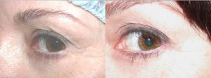
Below is an example of a lateral canthal web repair. On the left the web is present on a patient who had blepharoplasty surgery. Note how the webbed skin blocks vision on side gaze. On the right is the patient 6 months after repair. I would be happy to see you, or anyone you know who needs assessment and treatment for a canthal web, or for any revisional eyelid procedure


