
Lacrimal Gland Repair
One of the lesser-known parts of the eyes are the lacrimal glands. These glands help with tear production. In some cases, the lacrimal gland can become dislocated and create a bulging appearance at the outer corner of the upper eyelid. To correct the appearance of a prolapsed lacrimal gland, you will need to seek out an experienced oculoplastic surgeon, like Dr. Guy Massry in Beverly Hills, who specializes in lacrimal gland repair.
What is a prolapsed lacrimal gland?
The lacrimal glands are part of your orbital anatomy. They are located in the upper outer corner of the eye (lacrimal fossa) and are responsible for producing tears. Lacrimal gland prolapse (or a dislocated lacrimal gland) results in a bulging appearance to the outer part of the upper eyelid. In addition to an unattractive appearance, some patients will feel pressure on the globe and may experience discomfort. A prolapsed lacrimal gland can occur with aging or after trauma.
What is lacrimal gland repair?
In chapter 10 of Dr. Massry’s latest textbook “Master Techniques in Facial Rejuvenation” Second Edition, he discusses the management of lacrimal gland prolapse as an adjunct to upper blepharoplasty. Lacrimal gland prolapse has been reported in 15% of all upper eyelid blepharoplasty patients and in up to 60% of patients who are undergoing functional upper blepharoplasty.
Suture suspension of the lacrimal gland is a direct way to address moderate to severe lacrimal gland prolapse. The prolapsed gland is identified through the standard upper eyelid blepharoplasty incision. The lacrimal gland is pale in color with glandular elements and is distinctly different in appearance than the closely located central fat pad.
To perform lacrimal gland repositioning, Dr. Massry uses a 5-0 polypropylene suture on a P-3 needle to pass through the lacrimal gland capsule. The suture is then secured to the anterior tip of the lacrimal gland fossa periosteum at the superior orbital rim. He finally ties the suture, which repositions the gland into the fossa. This technique allows the eyelid to close normally.

Lacrimal Gland Repair
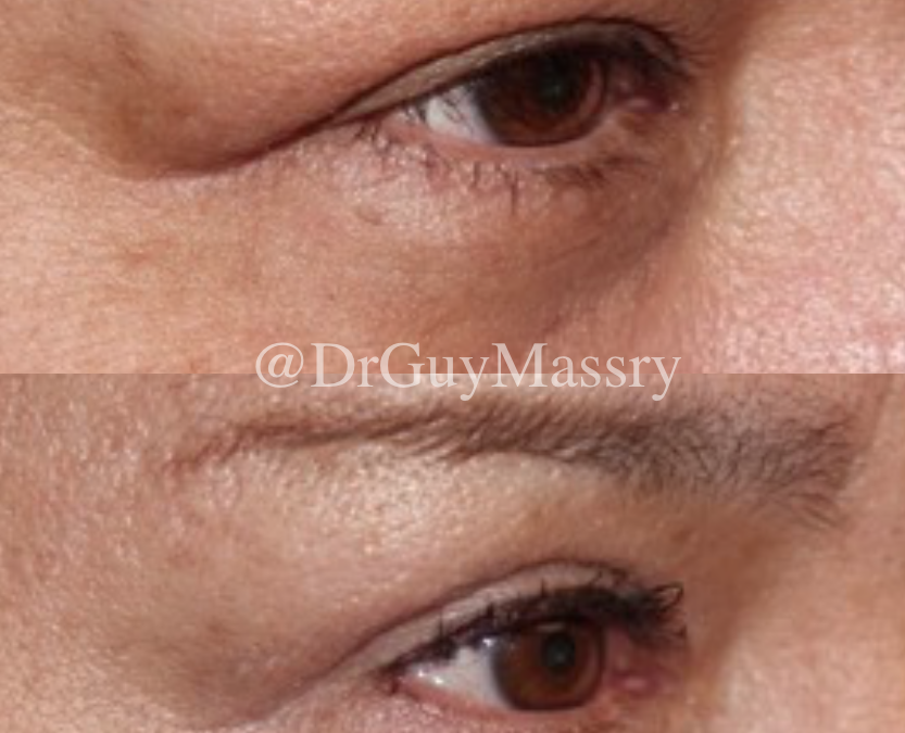
View examples of patients before and after prolapsed lacrimal gland repair. As you can see in the before pictures, there is a bulge in the outer corner of the eyelid due to the dislocated lacrimal gland. After the lacrimal gland is repositioned to its correct position, the patient looks much more youthful.
Videos
These videos provide a look into what to expect during your lacrimal gland surgery.
For answers to frequently asked questions, head over to our What to Expect page.